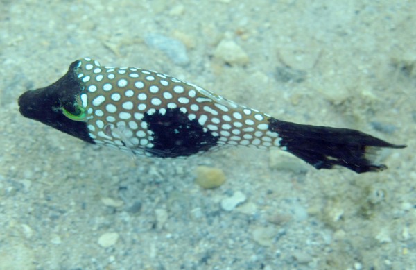
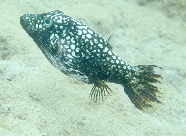
LIHU‘E — Something is causing Hawaiian white-spotted toby fish along Kaua‘i’s North Shore to develop skin ulcers and inflammation. But after more than a month of studying collected fish, scientists are no closer to having an answer. A diagnostic report released
LIHU‘E — Something is causing Hawaiian white-spotted toby fish along Kaua‘i’s North Shore to develop skin ulcers and inflammation. But after more than a month of studying collected fish, scientists are no closer to having an answer.
A diagnostic report released Wednesday by the U.S. Geological Survey details Dr. Theirry Work’s findings — or lack of.
“There’s clearly some sort of cause … but I wasn’t able to get an answer,” Work, a wildlife disease specialist for the USGS’s Honolulu field office, said over the phone Wednesday, shortly after the report was sent out. “It’s a more complicated problem.”
Between Nov. 27 and 29, Work, along with Dr. Greta Aeby — a coral expert with the Hawai‘i Institute of Marine Biology at the University of Hawai‘i — collected a total of seven toby fish from the waters off ‘Anini Beach. The fish, which have been showing up with black skin discolorations and unusual lesions, were collected and examined as part of their ongoing study of a cyanobacterial/fungal disease killing coral at ‘Anini, Makua and Hanalei.
While Work has not been able to pinpoint the cause, he said he has clarified that the situation is “truly abnormal,” as well as ruled out several potential sources.
“I’ve definitely ruled out things like bacteria, fungi or parasites,” he said. “The bottom line is to sort this out is going to take a little more footwork and shoe leather.”
In the report, Work writes that all of the collected fish had varying degrees of distinct, shapeless black discoloration, mainly affecting the animals’ nose or tail fin, and in some cases, their entire body.
“It’s a chronic inflammatory response,” Work said. “I’m thinking some sort of virus or some sort of contact irritant.”
Whatever the cause, Work said it is leading to the skin “ulcerating away.”
“Rarely, mild inflammation or cell death was seen,” he wrote in the report. “One fish had marked inflammation of the liver, two had varying degrees of worm infestation in the liver and two had bacterial infections in the gills.”
While the skin discolorations are abnormal, Work does not know whether there is a relationship between that condition and other problems found in the fish, including liver inflammation and gill infections.
Ultimately, the answer is “not something that’s going to be sorted out over night,” he said.
While similar skin problems have been seen in toby fish in O‘ahu and Kaua‘i’s South Shore, no microscopic study was ever done, according to the report.
One thing that Work said he found “curious” is that the ulcers are affecting the tobies’ noses and tails.
“Why is it at both ends of the fish?” he asked.
Looking forward, Work wrote that it would be informative to quantify the percent of fish affected — by what he described as “an easily detectable condition” — and determine if the ulcers and inflammation is somehow related to the condition of the reefs.
Additionally, he said he hopes to experiment with affected fish in a captive setting.
“If you take a fish out of the habitat and put him in an aquarium, does it go away?” he questioned.
Work concluded Wednesday’s report by writing, “Sorting this out would require further study.”
Since September, Work and Aeby have made several trips to Kaua‘i to photograph infected coral colonies, document the disease’s progression and collect samples for further study.
Work’s initial findings were outlined in a Nov. 21 diagnostic report, in which he called the outbreak an epidemic and said it is the first cyanobacterial disease documented in Hawaiian corals.
• Chris D’Angelo, lifestyle writer, can be reached at 245-3681 (ext. 241) or lifestyle@thegardenisland.com.


